What are Dentigerous Cysts?
A dentigerous cyst can occur when a tooth fails to erupt normally as the animal grows, often due to the tooth being impacted or embedded. This occurs more frequently in brachycephalic breeds, possibly due to the shape of the skull or the overcrowding of teeth which can impede normal tooth eruption. When a dentigerous cyst occurs, it will arise from the enamel epithelium and surround the crown of the unerupted tooth. Often the cyst is fluid-filled and can be soft or firm depending on the presence of bone involvement. In some cases, dentigerous cysts can lead to pathologic fractures as they grow and weaken the surrounding bone. While other types of odontogenic cysts can occur in dogs, dentigerous cysts are the most common.
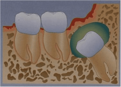
Figure 1. © studyblue.com
Clinical Signs of a Dentigerous Cyst
Dentigerous cysts often have no clinical signs and may go undetected on routine oral examinations. Thus, any animal with a missing or unerupted tooth should be carefully evaluated for the presence of a dentigerous cyst. When a cyst occurs, the area is typically not painful as long as the cyst does not become infected. In some dogs, the tissues overlying the cyst will appear normal, while in others a visible oral swelling may be present. In most cases, there will be no other noticeable clinical signs to indicate a problem. Because of this, dentigerous cysts may go undiagnosed until they reach considerable size.
In severe cases, the dentigerous cyst can grow large enough to damage the adjacent cortical bone, leading to facial asymmetry, pathologic fractures, and a visible oral mass. These larger, more severe lesions are typically associated with a later diagnosis.
Any unerupted tooth can develop a dentigerous cyst, but the condition most commonly involves the mandibular first premolar, the mandibular third molar, the mandibular incisors, or the canine teeth. Any unexplained missing teeth in these areas should be considered cause for further evaluation.
Diagnosis of Dentigerous Cyst
Dentigerous cysts often present as simply an unexplained missing tooth, so a complete oral exam under general anesthesia is the first step in identifying the problem. Fine needle aspirates of the cyst may yield fluid that varies from serosanguinous to yellow or brown, but this is often diagnostically unrewarding. Dental radiography should be performed to evaluate any missing or unerupted teeth, and most dentigerous cysts can be definitively diagnosed based on the radiography findings. Although radiographic findings are often pathognomonic for dentigerous cyst, it is also recommended that any excised tissues be submitted for histopathology to rule out other causes of oral swelling, such as abscess, granuloma, and neoplasia. While the vast majority of dentigerous cysts are benign, neoplastic changes in the cyst wall are possible.
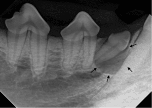
Figure 2. Typical radiographic appearance of a dentigerous cyst (black arrows)
Treatment of Dentigerous Cyst
Fortunately, treatment is fairly simple when the dentigerous cyst is identified early. The treatment of choice is extraction of the unerupted tooth and surgical excision of all cyst tissues. In cases where the cyst has become particularly large, damage to the surrounding tissues may result in bone loss, necessitating a bone graft. In these cases, follow up radiographic monitoring of the cortical bone is recommended to ensure normal healing occurs after the cyst is removed. As discussed above, it is recommended that excised tissues be submitted for histopathology to confirm the diagnosis of dentigerous cyst.
Any unerupted tooth has the potential to form a dentigerous cyst, so it is recommended that these teeth be extracted early to reduce complications. If extraction is not performed, close monitoring of the unerupted teeth is necessary to identify and address cysts early.
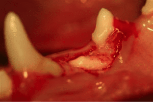
Figure 3. Surgical approach to extraction of an unerupted first premolar tooth
Outcomes
Surgical removal of the dentigerous cyst and extraction of the unerupted tooth is curative in most cases, and the prognosis is excellent. If excision is incomplete and cyst remnants are left in the oral cavity, the cyst may recur, necessitating further surgical intervention. In cases where the cyst has become particularly large, further treatment to address bone loss and pathologic fractures may be necessary.
Summary
Dentigerous cyst is a fairly common dental condition in dogs that is fortunately easy to treat if diagnosed early. Because the condition can be subtle and often presents with no clinical signs, any missing or unerupted teeth beyond the normal period of eruption should be thoroughly evaluated. Particularly close observation is necessary in brachycephalic breeds, in which skull shape and crowding of teeth may predispose the patient to the development of dentigerous cysts. In most cases, surgical excision of the cyst and extraction of associated tooth is curative.